
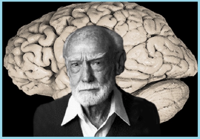
As a neurobiologist in the 1940s, Nobel laureate Roger Sperry was responsible for some of the most groundbreaking operations in medical history. All of his patients were newts. Sperry began by carefully removing the eyes of newts using jeweler’s forceps. He turned them around by 90 degrees and pushed them back into place.
After two days of rest, Sperry continued the operation on the newts. To do this, he cut into the top of each newt’s mouth and opened the sheath protecting the optic nerve, which carries visual information from the eyes to the brain. He removed the nerve from its sheath, snipped it in half, and replaced the two halves.
Roger Sperry Was Looking For a Cure For Which Disease
It’s safe to assume that any human subjected to Sperry’s brutal procedure would have been left permanently blind. The ability of newts to regenerate nerves, however, is truly remarkable. After a month, those who were treated by Sperry had restored vision.
There “was not a fuzzy muddle” in their perception, he penned. One of the newts lunged quickly as he dangled a bait in front of it. The animal lunged in an unusual direction, looking up when the bait was held low and down when it was held high. In other words, Sperry had completely flipped the newt’s world on its head.
The experiment demonstrated that neurons, the cells that make up the nervous system, are capable of an impressive degree of self-wiring. Dendrites extend from neurons to take in information, and axons extend from them to send the information on to other neurons. In instance, axons may traverse incredible lengths while yet pinpointing their destination with incredible accuracy. They are capable of winding their way through the maze of neurons in the brain, forging synapses with only the most compatible partners despite the presence of billions of other neurons on their path.
60 Years of Study
Sperry’s newts’ retinal neurons regrow axons that connect to those in the brain’s visual cortex. Eye axons apparently maintained their connections with the same regions of the brain after surgery. Since the eyes had been rotated during surgery, but the neural connections that they created unfolded normally, the only change was that the eyes delivered inverted images after the procedure.
After 60 years of study, it is now obvious that Sperry’s newts were not exceptional. In every living thing, the nerve cells precisely and reliably wire themselves together to form a functioning neurological system. In humans, this process initiates during gestation, when the brain’s initial neurons are formed. Their longest axons reach all the way from the tips of the toes to the bottom of the spine. Some neurons continue to wire themselves long after brain development is complete, as nerves repair from minor injuries and axons form new connections when we acquire new abilities.
Last Words
Many things go wrong with our bodies and brains when neurons fail to link properly. Duane syndrome affects around 1 in 1,000 newborns; it is characterised by misfiring axons in the nerves that normally regulate the eye muscles. Sometimes axons fail to properly direct their growth, and they end up on the outside of the eye rather than the inside, where they belong (in the muscle of the inner corner of the eye).
People with this disease have trouble turning their eyes in because the inner corner muscle contracts when they make an inward movement. The same signal, however, is sent to the muscle on the periphery. When both muscles contract simultaneously, the eye is yanked back into place.


")


